Veterinary Services

Pet Orthopedics in Kingsport, TN

Learn more about our orthopedic surgery services below!
Kingsport hospice end of life care
Veterinary Services

Pet Orthopedics at Kingsport Veterinary Hospital.

Orthopedic conditions affect your pet’s skeletal system and limbs, including bones, joints, muscles, tendons, and ligaments. These injuries can have a profound impact on your pet’s mood and quality of life.
Kingsport Vet Hospital - pet wellness exams

My dog needs surgery TPLO versus Lateral Suture

Dogs have a cranial and caudal cruciate ligament, like a human’s anterior (ACL) and posterior ligaments. The cranial cruciate ligament (CCL) is the one most commonly torn and is one of the major stabilizers in the knee. Rupturing the CCL is one of the most common causes of lameness in the rear legs of dogs. Veterinarians often perform tibial plateau leveling osteotomy (TPLO) surgery to fix the tear.

READ MORE

Are their risk factors for cruciate tears?

Almost all breeds of dogs and cats are at risk, but some breeds are predisposed to a CCL tear. It is seen most often in Labs, Golden Retrievers, German Shepherds, Boxers, Pit Bulls, American Bulldogs, Mastiffs, Rottweilers, and Newfoundlands.

Poor physical condition and obesity predispose dogs to tearing the ligament, but fit dogs can also get a tear. Ligament tears in dogs are different than in humans. In dogs, the ligament starts to degrade and becomes weak over time whereas humans usually tear during sports. Since one ligament degrades and fails, 40-60% of dogs tear the other knee within two years of the first injury.

Injury can occur at any age but is most common

What are the signs of CCL tears?

Dogs can present in a variety of ways. Some dogs will suddenly become lame on the leg and may hold it in the air. Some dogs will have a mild lameness for months that will get better and worse over time. Other dogs will have the intermittent mild lameness and then suddenly become very lame.

However, if both knees are torn at the same time (which occurs in 30% of dogs), they can present unable to walk in the rear legs. Other signs of dogs with bilateral CCL tears can be difficulty going up stairs or jumping into the car, and difficulty sitting down and placing both legs out to the side (rather than sitting square).

How is a CCL tear diagnosed?

Physical examination is the most common method of diagnosis. The knee will feel thickened from the bones shifting back and forth since there is no ligament holding them together. They will have joint swelling, pain on hyperextension of the knee, and will often sit with the leg kicked out to the side.

The tibial thrust test and the cranial drawer test are the two main tests for instability in the knee.

  • For the tibial thrust test, the dog often stands (it is less stressful) and your veterinarian will hold the femur (thigh bone) stable while bending the foot. The tibia (shin bone) should not move, but if the cruciate is torn, then it will thrust forward. This happens because the top of the tibia is on a slope and the cruciate holds the femur on the slope. I make an analogy of a wagon being held on a hill by a rope. If the rope is cut, the wagon will roll down the hill. If the cruciate is torn, the femur will fall down the hill with every step they take (often crushing and tearing the meniscus (cartilage cushion in the joint).
  • The cranial drawer test involves holding the femur and the tibia and trying to move the tibia forwards. But in large strong dogs, it is very difficult to get cranial drawer without sedation.

Your veterinarian may also do an X-ray. Although you cannot see the ligament on an X-ray, there are classic changes seen on an X-ray such as swelling and arthritis. Dogs do not develop arthritis in the knee for no reason, and the most common cause is cruciate tears.

What are the surgical options for CCL tears?

There is no best surgery for cruciate tears, but most surgeons (and veterinary journals and research studies) agree that the TPLO is the best surgery for dogs. TPLO stands for tibial plateau leveling osteotomy. Another potential surgery is the lateral suture.

TPLO Surgery

Back to the wagon analogy for the best explanation. If we take the wagon and place it on a flat road, we don’t need a rope to hold it stable. With the TPLO, the surgeon cuts the back part of the tibia with a special circular saw and rotates the piece of bone so the top of the tibia is almost flat. Then the surgeon repairs the bone with a special bone plate to hold it in place while it heals. Once the bone is healed in the “flat road” position, the tibia no longer thrusts forward when the dog walks and the knee is stabilized. The plate usually remains on the bone indefinitely, but it is no longer needed for support once the bone heals.

Learn More

Lateral Suture

This surgery involves placing a suture, which is usually strong nylon (similar to a strong fishing line), around the outside of the joint to hold the two bones together and prevent them from shifting. The problem with this surgery in large dogs is that there is a lot of force on the nylon and it tends to break and fail. Studies have also shown dogs are in more pain after a lateral suture and develop worse long-term arthritis. Many veterinarians reserve the lateral suture for small breed dogs.

Is TPLO surgery painful?

Using a saw to make a bone cut is different than a trauma causing the bone to break. There is usually not a great deal of swelling or bruising. The TPLO surgery is usually more comfortable than the lateral suture which feels very tight around the joint. Most dogs are using the leg within a few days post operatively, and they will often be more athletic after a TPLO than a lateral suture. Pain management is also critical with using epidurals, infusing the incision with a numbing agent that lasts for three days, and post-operative pain killers.

What is the long-term success with the TPLO?

Most data show that 90-95% of dogs will return to their previous activity after the TPLO. All dogs develop arthritis within a few weeks of the ligament tearing due to inflammation, but once the knee is stabilized, this minimizes the arthritis. Dogs will improve up to six months after the surgery as that is how long it takes for them to build muscle strength and for the swelling to improve in the joint. Even with the arthritis, most dogs will only have occasional stiff days after strenuous play or a long rest, but quickly work out of it. Our surgeons have seen dogs going back to work on the Police force and performing agility after a TPLO surgery. However, it does take rehabilitation and muscle strengthening to achieve the best results.

What are the most common complications of a TPLO?

The two most common complications are infection and implant failure/fracture of the bone. Infection is the most common with studies showing an infection rate from 8-17%. The most common reason for infection is that dogs can lick the incision because the E-collar is removed.

If they lick the incision and an infection forms, it can move to cover the plate as there is not a lot of muscle or tissue over the plate. When that happens, it causes a slime coating over the plate that antibiotics cannot penetrate and kill permanently. If an infection forms, the bone usually heals fine, but the implant is often removed to resolve the infection. Fracture/failure is possible, but this is less common with the new Locking Plates and Screws and if the dog is kept restricted and there is no jumping or strenuous play.

How does my dog recover after TPLO surgery?

You should confine your dog to controlled leash walks for six to eight weeks post operatively while the bone heals. They are allowed to walk on the leg, but no jumping, furniture climbing, or running. They should be confined to a crate or very small room while they are healing. We recommend you keep them on a short leash so they can be controlled when they are out of the crate, but still feel a part of the family. Often sedatives prescribed to help keep calm and quiet.

Rehabilitation is recommended to build muscle strength and decrease swelling. Rehab is veterinarian prescribed for home and is prescribed based on healing. 

Help em up Harness. We will require a harness for dogs that cant be picked up all the time. Click the button below for more information about the harness.

HARNESS INFORMATION

Kingsport Vet Hospital - pet wellness exams

Lateral Cruciate Suture Repair

Extracapsular suture repair is a leading option for the treatment of unstable stifles due to cranial cruciate ligament insufficiency. Lateral suture repair for dogs has been around for decades and has helped tens of thousands of dogs. Although osteotomy techniques like TPLO and TTA have become the standard of care for large breed and athlete dogs, the extracapsular repair has remained a very useful technique that veterinary surgeons can perform in your hospital when referral is not an option.

READ MORE

This procedure has changed over the years to address the weaknesses of the original technique. The development of a tensioning and crimping system with nylon leader line was a quantum improvement over the original hand-tying system. Ongoing improvements have focused on increasing the strength of the implanted material, limiting its stretch, increasing the strength of the anchoring points, and maximizing isometry so the suture is uniformly tight during the entire range of motion.

New systems for this repair have incorporated these new features. Remember, however, that all these techniques are still mechanically extracapsular repairs. This means that they mechanically prevent cranial subluxation and nearly eliminate the normal internal rotation of the tibia. Most of the newer implants, whether a suture, tape, or loop, are made of ultra-high molecular weight polyethylene. This material is much stronger than nylon leader line and does not stretch. The anchoring points are bone tunnels with buttons or interference screws, screw-in anchors with holes or flanges, or screw-in anchors with the material already attached.

The implants’ placement counters the force of subluxation from the isometric points on the distal femur and proximal tibia. The Everost OrthoZIP system of lateral suture repair for dogs was developed by the same bioengineer who created the original nylon leader line tensioning and crimping system. This system employs a self-tightening, ultra-high-molecular-weight polyethylene loop that is placed over titanium anchors with a cancellous thread profile and a “sewing-bobbin” shaped head to retain the loop. After arthrotomy, inspection, and treatment of any meniscal injuries, the anchor points are identified, pre-drilled, and the anchors placed. The loop goes over the anchors, which we tighten by pulling on the two free ends. The loop maintains the tension without any knotting or crimping. Several sizes are available to cover a full range of patient sizes.

Kingsport Vet Hospital - pet wellness exams

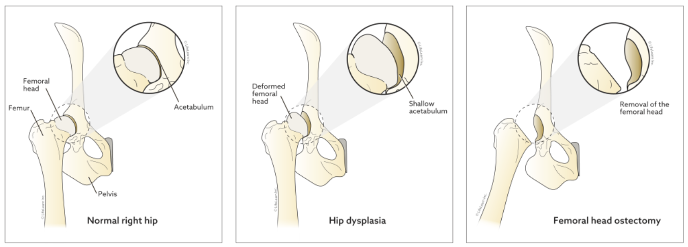
Femoral Head Ostectomy (FHO)

What is FHO surgery?

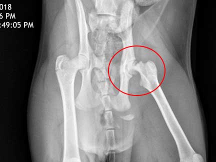
An FHO, or femoral head ostectomy, is a surgical procedure that aims to restore pain-free mobility to a diseased or damaged hip by removing the head and neck of the femur (the long leg bone or thighbone).

How does an FHO change the hip?

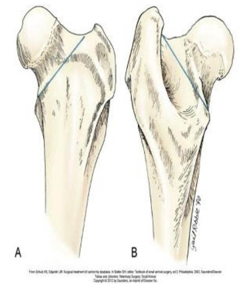
The normal hip is a ball-and-socket joint. The acetabulum, which is a part of the pelvis, composes the socket of the joint. The head of the femur, a projection from the long bone located between the hip and the knee, composes the ball that fits within the socket. The head of the femur fits within the acetabulum, allowing the hip to move freely in all directions.

READ MORE

Is my dog a good candidate for FHO?

This procedure is primarily recommended for small dogs (under approximately 45 pounds) and cats, especially those who are at a healthy weight. The false joint that is created in an FHO works very well to support the weight of small animals but may be less effective in large-breed dogs. There are exceptions, however, and veterinarians may recommend an FHO for a dog over 50 pounds if the specifics of the case dictate that doing so would be appropriate.

“Active dogs often experience better results with FHO than less active dogs.”

Active dogs often experience better results with FHO than less active dogs. The muscle mass that has been built up through activity helps to stabilize the joint, allowing the dog to regain pain-free mobility more quickly than inactive pets. Inactive dogs have less muscle mass around the joint, making the joint less stable post-operatively and leading to longer recovery times.

Why is FHO performed?

The primary goal of an FHO is to remove bone-on-bone contact, restoring pain-free mobility. The most common reasons for FHO include:

Fractures involving the hip. When a fracture involves the hip joint and cannot be repaired surgically (either due to patient considerations or financial considerations for the owner), an FHO may provide the best option for pain-free mobility.

Hip luxation/dislocation (associated with trauma or severe hip dysplasia). In some cases, a hip that is out of the socket cannot be replaced with manipulation or other medical means. Surgical repair of hip luxations can be costly and is not always successful, so many dog owners elect FHO for small dogs with hip luxation.

“The primary goal of an FHO is to remove bone-on-bone contact, restoring pain-free mobility.”

Severe arthritis of the hip. In chronic, end-stage arthritis, the cartilage that protects both the head of the femur and the acetabulum can become eroded away, leading to painful bone-on-bone grating whenever the hip is moved. Performing an FHO can remove this point of contact and alleviate pain.

Legg-Perthes disease (also known as avascular necrosis of the femoral head). This uncommon condition, most frequently seen in miniature and toy breed dogs, causes the bone within the femoral head to begin to die at an early age. The bone collapses due to these degenerative changes, leading to severe pain. Removing the femoral head via FHO removes the source of pain for the dog.

What can I expect on the day of surgery?

This surgery is performed under general anesthesia. In most situations, you will take your dog to the veterinary clinic early in the morning on the day of surgery. Your veterinarian will likely instruct you to withhold food the morning of surgery to prevent vomiting that may occur under anesthesia.

After surgery, your dog will remain in the hospital for several hours to several days depending on the specific circumstances of his health and his surgery. When you pick him up from the hospital, your dog probably will not be bearing any weight on the leg that had surgery.

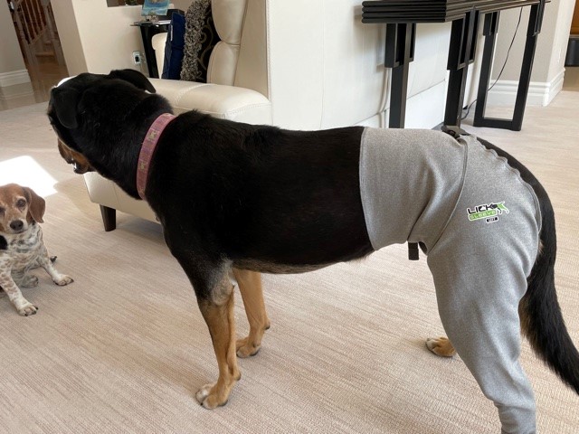
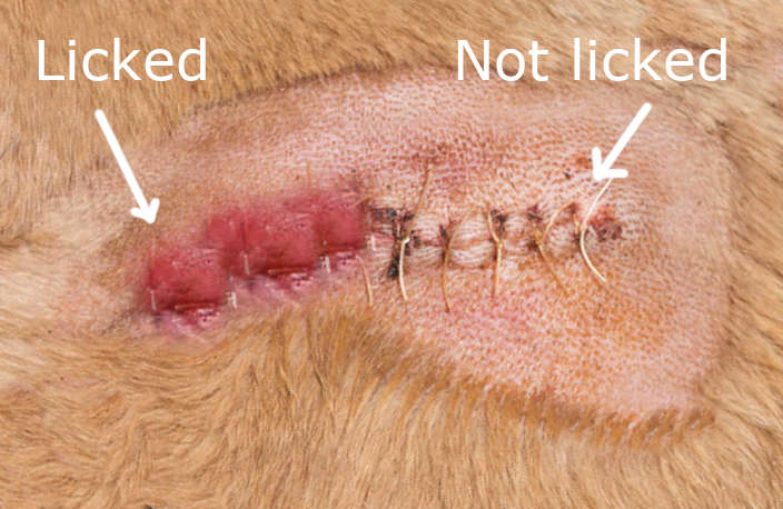
An incision will be visible in the area of the hip, and this incision may or may not have visible external sutures. Some veterinarians use dissolving sutures that are placed under the skin. Your dog will likely be wearing an Elizabethan collar (cone) to prevent licking at the surgical site.

What care will my dog need after FHO surgery?

Care varies based on the needs of the specific patient but, in general, the post-operative recovery can be divided into two phases. In the first several days post-operatively, your dog will be healing from the surgical procedure. Because bones and muscles are cut during this procedure, the focus during this period will be on pain control. Please give all medications as prescribed by your veterinarian. Moist heat may also be recommended during this period to provide comfort and decrease stiffness. Your veterinarian may also recommend laser therapy to reduce inflammation and encourage healing.

Your veterinarian may recommend activity restrictions during the first several days after surgery. If this is the case, confine your dog to a crate or a small room within the house, with only very brief leash walks outside to eliminate. If your dog will tolerate it, you can attempt passive range-of-motion (PROM) exercises during this period, gently moving the hip forward and backward through its range of motion. This should not be performed, however, if it causes pain for your dog.

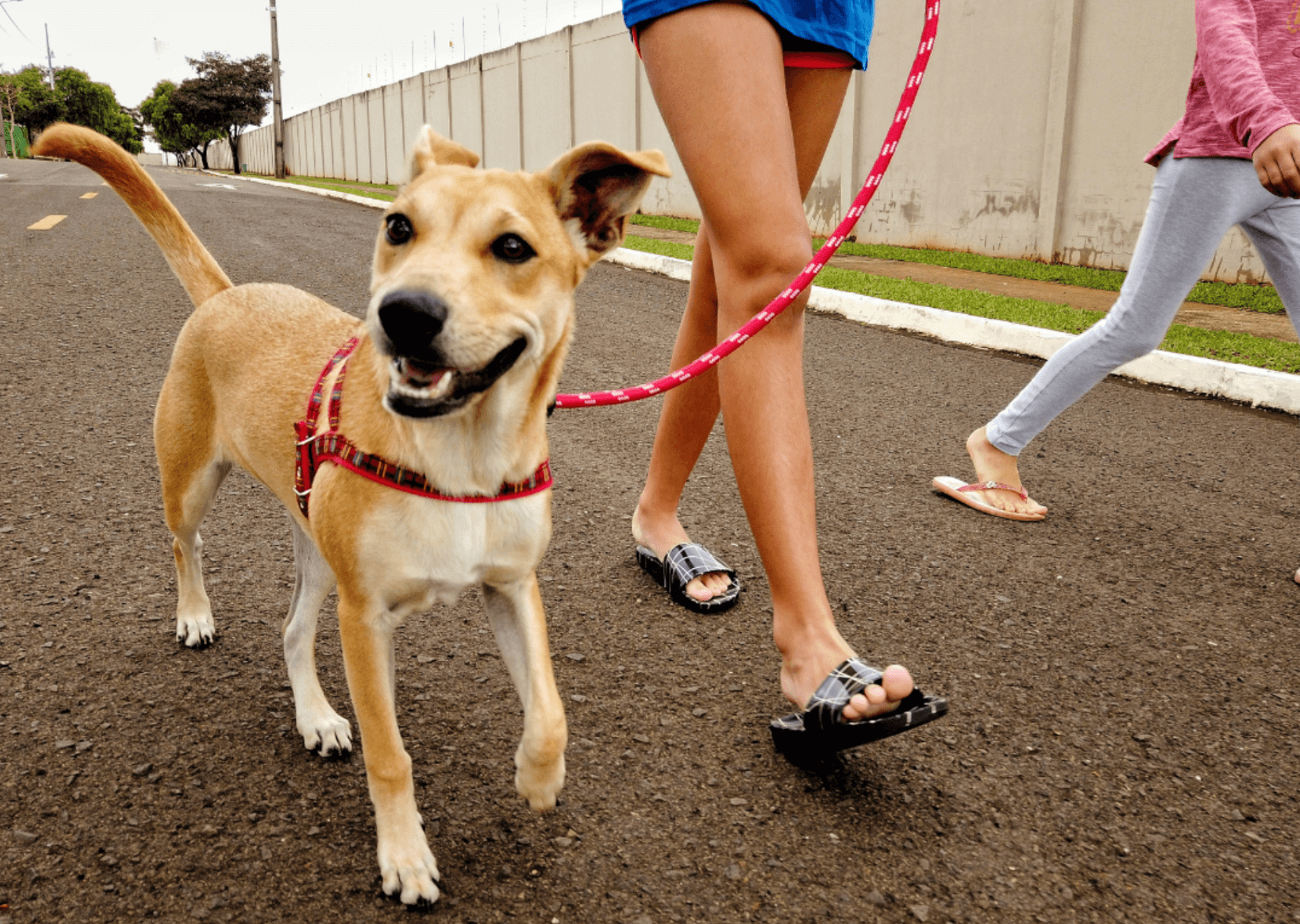
Your veterinarian will likely recommend introducing more physical activity approximately one week after surgery. During this phase of recovery, the focus shifts to rebuilding muscle mass and strength. Keeping your dog mobile will help keep the scar tissue within the false joint from forming too tightly, allowing your dog to remain flexible. Good exercises during this period include walking (especially up flights of stairs), holding the front portion of your dog’s body in the air while allowing him to ‘walk’ on his hind legs, and walking through water. Walking should be slow to encourage your dog to bear weight on the affected leg; when running, your dog will be more tempted to carry the affected leg.

“Keeping your dog mobile will help keep the scar tissue within the false joint from forming too tightly, allowing your dog to remain flexible.”

In the first 30 days after surgery, it is important to avoid rough play or any activity that encourages sudden twists and turns. These high-impact motions will slow the healing that is occurring within the joint and muscles.

Most dogs will show signs of complete recovery approximately six weeks post-operatively. At this point, your dog can resume his regular activities. Healing may be more rapid in dogs that had normal function up until shortly before the FHO (i.e., in the case of a dog that had a sudden, traumatic injury to the hip) and may be slower in dogs with longstanding, chronic issues (because these chronic issues often lead to muscle atrophy, which takes time to resolve).

If your dog is not showing significant improvement by six weeks post-operatively, you may want to consider a formal rehabilitation or physical therapy program. Ask your veterinarian for recommendations if your dog is still having difficulties at or after six weeks.

What is the prognosis after FHO surgery?

Most dogs recover fully after FHO surgery and regain essentially normal function of the affected leg. Although the leg may have a slightly decreased range of motion or decreased limb length after surgery, these impacts are typically minimal and do not impact the pet’s quality of life.